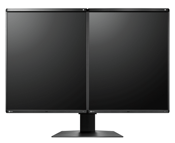
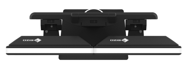
With a bezel only 7.5 mm in width – the world’s smallest bezel width for 5-megapixel monitors – the distance between the display areas of the two monitors is merely 15 mm. Moreover, the panel frame is only 2.5 mm above the screen, which means it sits nearly flush with the screen. This means viewers’ vision goes undisrupted when looking back and forth between the monitors.
The RX560 reproduces grayscale breast tomosynthesis images, mammograms, and color images from ultrasound scans or pathology examinations at the highest level of quality. The hybrid gamma PXL functionality ensures the highest levels of precision and reliability if color and monochrome images are displayed at the same time.
The RX560 MammoDuo saves a great deal of space. This solution saves 67 mm horizontally, 36 mm vertically, and 20.5 mm in depth, compared to conventional structures built from individual monitors with this resolution arranged next to one another. In sum, this means a 22% reduction of the total required surface. This frees up valuable space to make for a roomier working environment.
You can conveniently adjust the height, tilt, and rotation of the monitors with the dual stand, without creating gaps between the monitors.
Narrow black frontal bezels make this device ideal for use in dark environments. They make it easy to visually concentrate on the display. Meanwhile, a white bezel at the sides of the monitors creates a fresh, clean look.
The ‘point and focus’ functionality allows you to quickly select and focus on relevant image areas using your mouse or keyboard. The brightness and grayscale value of certain points on the screen can be adapted, if necessary, in order to optimize the display.
Mammography is increasingly being combined with ultrasound scans in early breast cancer detection, particularly for patients with a high breast density. Moreover, in cases of suspected breast cancer, additional methods such as biopsies, breast MRIs, and computer tomography are used.
The RadiForce RX560 is the world’s first medical monitor that uses an LCD on an LTPS (low-temperature polysilicon) basis. This allows the color monitor to achieve a brightness of up to 1100 cd/m2, comparable to that of a monochrome monitor. As a result, the RX560 is able to display high-resolution breast tomosynthesis images as well as mammograms with deep, non-faded black tones as well as color images from ultrasound scans and pathology examinations.
A high contrast ration of 1500:1, close to that of a monochrome monitor, means that deep black tones can also be reproduced without a ‘washing out’ effect.
The hybrid gamma PXL functionality automatically differentiates between monochrome and color images, pixel by pixel. This creates a hybrid display on which each pixel is displayed with the ideal tone value. In turn, this achieves a greater degree of precision and reliability than for conventional planar detection methods.
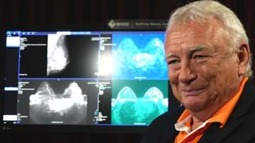
The RX560 reproduces sophisticated monochrome images from breast tomography or mammography as reliably as color images from breast MRIs or CTs, ultrasound scans, and pathology. In practical application, this means a significant increase in efficiency, since images from various imaging methods can be observed on a single monitor.
Thanks to the signal input and output, you can link several RadiForce monitors through their DisplayPort interface. This means that you can realize multi-monitor solutions with the greatest of ease – without laborious and excessive cabling.
LCD panels with a high brightness level tend to have more blurry image rendering thanks to over-framing than would be possible in comparison with an acquired exposure. Therefore, EIZO offers blur reduction anchored in monitor hardware. It retrieves details lost in the contours on the screen, meaning that the image is rendered as clearly as possible.
The monitor has a pixel width of 0.165 mm and thereby reproduces even, high-resolution, sharp, and high-depth images without any kind of granularity.
The display properties, in particular brightness and contrast, are suited to the creation of image rendering systems compliant with DIN 6868-157. The DICOM GSDF characteristic is already precisely configured in the factory. This means that grayscales are consistent, which is vital for diagnostics.
The monitor meets the strictest medical, safety, and EMC emission standards. EIZO’s QC software RadiCS enables you to perform brightness, grayscale and uniformity checks that comply with AAPM TG18, DIN 6868-157, and other QC standards.
The precise calibration of white point and tone value characteristic curve is provided by an integrated front sensor (IFS). This measures the brightness and grayscales and calibrates the monitor autonomously according to the DICOM standard. The sensor works automatically, without restricting the field of vision of the monitor. You can save the costs, time, and effort of maintenance and rely on a consistently balanced image quality.
Precise color reproduction is controlled via a 12-bit look-up table (LUT). A maximum of 10-bit resolution, or up to one billion hues, is available via DisplayPort. This ensures flawless color reproduction of MRI, ultrasound, and pathology images. As such, the recording curve and microstructures required for diagnosis can be precisely detected.
A sensor for the backlight permanently determines the luminance of the monitor. The benefit: The defined and calibrated values are rendered exactly just seconds after the monitor is turned on and remain constant during the entire period of use. The sensor is invisibly integrated in the monitor.
The monitor shines thanks to its high color purity and uniform illumination. This is down to the Digital Uniformity Equalizer (DUE), which corrects imbalances automatically, pixel by pixel. Gray and color tones of radiological and other medical images are correctly rendered over the entire display. This is vital for diagnostics.
The CAL Switch functionality enables you to select from among many different display modes for different modalities, such as mammography, breast MRIs, ultrasound, or pathology examinations, without having to recalibrate each time.
Shipped with the monitor, the RadiCS LE software allows users to set modes in such a way that the optimal observation conditions are automatically activated, either via mouse click or through the monitor’s display mode.
Thanks to the presence sensor, you can save electricity and help protect the environment. The sensor registers whether someone is sitting in front of the screen or not. As soon as the person leaves the workstation, the monitor turns off automatically. When the person comes back, it turns back on – fully automatically, without touching the mouse or keyboard. It is always ready for use without a waiting period.
EIZO is convinced of the quality of its products. The warranty for the monitors, therefore, also covers the brightness stability.
The optional EIZO RadiCS software to secure image quality enables extensive maintenance and testing of monitors and includes calibration, acceptance and constancy testing, and the archiving of all areas. If you are working on multiple stations, the use of the RadiNET Pro is recommended. This can be used to centrally control the calibration of all monitors, including data history. This saves you a significant amount of time and ensures consistently high image quality across the entire setup. The basic version RadiCS LE is already included with the RadiForce GX, RX, and MX/MS models.
Learn more about the RadiCS application classes
Learn more about RadiCS LE software (included in the delivery)
Learn more about RadiCS software (optionally available)
Learn more about RadiNet Pro software (optionally available)
The EIZO MED-XN91 graphics card supports the properties, functions, and settings of the RadiForce RX560-MD optimally. It enables precise diagnostics and can control several monitors simultaneously. EIZO offers technical support and a warranty service for all graphics cards. Therefore, we recommend using EIZO graphics cards.
EIZO offers a brand-new, easy-to-operate comfort light for radiologists who work in dark diagnosis rooms. The soft illuminance in the background of the screen reduces the strain on the eyes that frequently occurs due to constant light-dark changes between bright screens and objects in a dark environment.
- Two 5-megapixel color LCD screens with consistently high and stable brightness for clear mammogram imaging
- Clear perceptibility of microstructures through high contrast and Sharpness Recovery technology
- Palette with 68 billion hues for precise color reproduction (10-bit resolution max.)
- Hybrid gamma PXL functionality for precise display, down to the pixel, of grayscale and color images with the required luminance characteristic curve
- Homogenous display surface with automatic luminance distribution control (Digital Uniformity Equalizer)
- Set up for calibration, acceptance, and consistency testing in accordance with DIN 6868-157 and QS-RL
- Effortless quality control and built-in calibration sensor
- Light sensor to measure ambient light at the diagnostic station
- Compact dual-screen solution through a shared stand with narrow bezels and ergonomic design
| Model Variations | RX560-BK-MD: Anti-Glare coating, two screens, with dual stand, black RX560-ARBK-MD: Anti-Reflection coating, two screens, with dual stand, black RX560-BK: Anti-Glare coating, one screen, with stand, black RX560-ARBK: Anti-Reflection coating, one screen, with stand, black |
|---|---|
| Panel | |
| Type | Color (IPS) |
| Backlight | LED |
| Size | 54.1 cm / 21.3" |
| Native Resolution | 2048 x 2560 (4:5 aspect ratio) |
| Viewable Image Size (H x V) | 337.9 x 422.4 mm |
| Pixel Pitch | 0.165 x 0.165 mm |
| Display Colors | 10-bit colors (DisplayPort): 1.07 billion (maximum) colors 8-bit colors: 16.77 million from a palette of 543 billion colors |
| Viewing Angles (H / V, typical) | 178° / 178° |
| Brightness (typical) | 1100 cd/m2 |
| Recommended Brightness for Calibration | 500 cd/m2 |
| Contrast Ratio (typical) | 1500:1 |
| Response Time (typical) | 12 ms (on / off) |
| Video Signals | |
| Input Terminals | DisplayPort, DVI-D (dual-link) |
| Output Terminals | DisplayPort (daisy chain) |
| Digital Scanning Frequency (H / V) | 31 - 135 kHz / 23 - 61 Hz |
| USB | |
| Upstream | USB 2.0: Type-B |
| Downstream | USB 2.0: Type-A x 2 |
| Power | |
| Power Requirements | AC 100 - 240 V: 50 / 60 Hz |
| Typical Power Consumption | 43 W |
| Maximum Power Consumption | 87 W |
| Power Save Mode | 1 W or less |
| Sensor | Backlight Sensor, Integrated Front Sensor, Presence Sensor, Ambient Light Sensor |
| Features & Functions | |
| Brightness Stabilization | Yes |
| Digital Uniformity Equalizer | Yes |
| Hybrid Gamma PXL | Yes |
| Preset Modes | CAL Switch |
| OSD Languages | English, German, French, Italian, Japanese, Simplified Chinese, Spanish, Swedish, Traditional Chinese |
| Physical Specifications | |
| Net Weight | RX560-MD, RX560-AR-MD: 17.3 kg RX560, RX560-AR: 8.1 kg |
| Net Weight (Without Stand) | 5.3 kg |
| Hole Spacing (VESA Standard) | 100 x 100 mm |
| Certifications & Standards (Please contact EIZO for the latest information.) | RX560, RX560-AR: CE (Medical Device), EN60601-1, ANSI/AAMI ES60601-1, CSA C22.2 No. 601-1, IEC60601-1, VCCI-B, FCC-B, CAN ICES-3 (B), RCM, RoHS, China RoHS, WEEE, CCC, EAC |
| FDA | 510(k) Clearance for Breast Tomosynthesis, Mammography, and General Radiography |
| Dedicated Software | |
| Monitor Quality Control Software RadiCS | Supported |
| Supplied Accessories (May vary by country. Please contact EIZO for details.) | |
| Signal Cables | RX560-MD, RX560-AR-MD: Dual Link DVI-D (3 m) x 2, DisplayPort (3 m) x 2, DisplayPort (1 m) RX560, RX560-AR: Dual Link DVI-D (3 m), DisplayPort (3 m) |
| Others | RX560-MD, RX560-AR-MD: AC power cords (3 m) x 2, USB cables (3 m) x 2, Utility Disk (RadiCS LE, PDF installation manual), instructions for use RX560, RX560-AR: AC power cord (3 m), USB cable (3 m), Utility Disk (RadiCS LE, PDF installation manual), instructions for use |
| Warranty | Five Years |
| Dimension Drawing |


